Mechanism and Kinetics of AMP-induced Pore formation in model membrane/biomembrane in Hypotonic Environment:
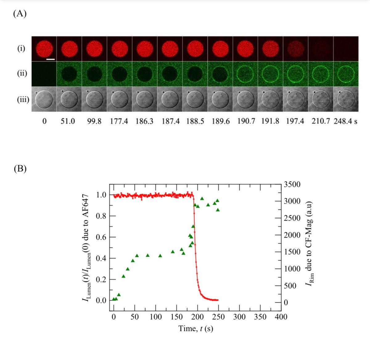
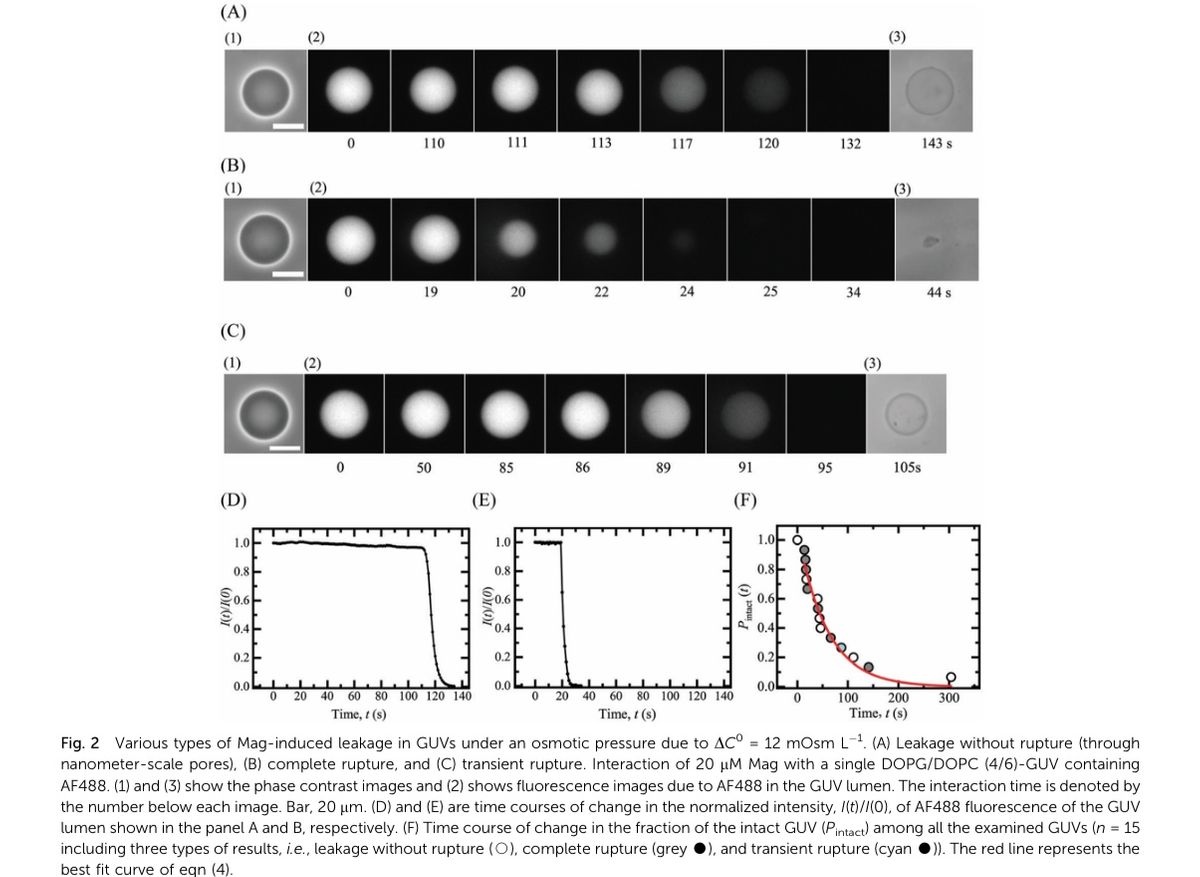
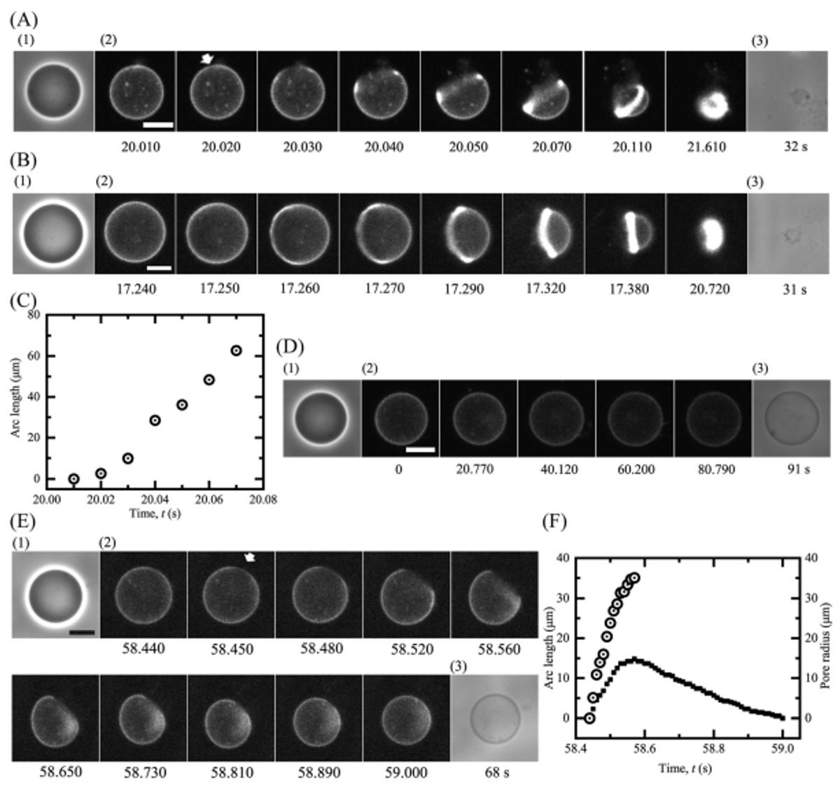
The AMP M2(synthesized, labeled, cleaved, purified and analyzed by us) was cationic in nature. In contrast, our model membrane (mimicking the Lipidic region of bacterial membrane) is negatively charged. So, there’s both electrostatic interaction (here, attraction) and hydrophobic interaction. When M2 binds in the outer leaflet of membrane and reaches the equilibrium, membrane tension induced in the inner leaflet as an effect of asymmetric peptide binding in membrane interface. As a result, pore forms in the membrane through which internal contents of Vesicles (including sucrose and water-soluble fluorescent probe) leak out as the efflux under positive osmotic pressure. Thus, lumen intensity of the GUV decreases, and phase-contrast also decreases greatly because of the minimizing the difference of refractive indices of internal and external environments of GUVs. Under positive osmotic pressure, a subset of total investigated GUVs ruptured due to the interaction of M2 with the Single GUVs. To elucidate M2-induced rupture of GUVs under osmotic pressure, I investigated the process of rupture of GUVs using High Speed Streaming and with high time-resolution and revealed the rupture of GUVs initiates from the pore formation. To reveal the Mechanism of pore formation event, I performed M2-induced area change in membrane using DIC (Differential Interference Contrast) microscopy. To reveal the location of M2 before and after pore formation, I used Confocal Laser Scanning Microscopy technoque.
Research Images


